The Angio Institute Research & Education Brain Health Program AngioNews/Media Joining Hands

When considering the purchase of any health or wellness device, today’s health professional and consumer are far more demanding of deeper answers about innovations. With high speed access to web research are at our fingertips, cutting through the jungle of marketing ads and product hype (to access proven science and technical data) is part of our info culture for the ‘smarter tech shopper’. Thanks to web research, we can make far better choices thanks to user reviews, company background checks, case studies or testimonials. HEALTH TECH REPORTER, an allied partner of the AngioFoundation presents a clinical perspective on health technologies from seasoned and unbiased testers and performance examiners to share their reviews for your added insight.

SCIENTIFIC REPORTING SUPPORTS
Validation & Claims
When considering the purchase of any health or wellness device, today’s consumer is more demanding of answers about what is on the market. With high speed access to web research are at our fingertips, cutting through the jungle of marketing ads and product hype (to access proven science and technical data) is part of the modern culture of the ‘smarter shopper’. With a little web research, we can make far better choices thanks to user reviews, company
background checks, case studies or testimonials.

Especially in the case of medical devices and health products, public interest for true quality and performance calls for a more critical eye, one that is not influenced by conventional sales and marketing methods. The value of a good review from AN OUTSIDE, INDEPENDENT TESTER (led by an uncompromised team) can offer a more confident understanding and technical credence about the product in question. It is the process of this type of ‘test drive’ that can offer valuable insight from actual clinical professionals. MEDTECH REVIEWS* aims to bring this unquiue evidence and SCIENCE based observation about the effects of the selected health innovations under review.


A MORE INVOLVED & UNBIASED TEST DRIVE
The AngioFoundation is one of the major non‐profit groups who supports the awareness about modern, alternative and NON‐INVASIVE INNOVATIONS. Through a network of clinical professionals and credentialed health advocates, the MedTech Reviews* program was formed to conduct case studies and research reviews on health related innovations. Combining multiple modalities of evaluation protocols (including the use of diagnostic imaging, behavioral studies and efficacy assessments), an unbiased focus group is designated for each project review to conduct a thorough user exam. Reports may include videos, imaging scans, recorded biometric tracking, user experience and other vital information to substantiating an intelligent review (and consumer activity). Our reviews are submitted for public access and digital distribution through our network of science news outlets. SCANNING TECH PERFORMANCE AND PHYSIOLOGICAL RESPONSE. This public education effort is lead in part by diagnostic imaging researchers and an evaluation team (assembled around each device). This test phase may be comprised of using a 3D Doppler Blood Flow Ultrasound Imaging to add to biometric logging and monitoring of a time‐based study on the effects of the device under review. This review can be accessed in the newsletter called JOURNAL FOR MODERN HEALING, a public blog‐style site where we present quantifiable scanning reports and interpretation analyses of all scanned readings. We provide this review to the public as part of an educational initiative to support beta testing with multiple modalities, valuation based on multiple opinions and use of non‐invasive imaging.

EVIDENCE-BASED PERFORMANCE REPORTS
The following are excerpts from actual pilot studies and performance reports of medical / therapeutic solutions:

•11/24 - Image Guided Approach to the Potency of PEMF on Prostate Hyperplasia (PDF report- official)
• 10/15 - Phase 1 Pilot Study of Exosome Topical Product (PDF report)
• 9/11/2023 - Performance Micro Study of (Aspen) Photobiomodulation (PDF report)
• 8/18 - Interventional Monitoring of Muscle Reaction to PEMF Induction (PDF official report)
• 6/1 - Mindray Sample Data/Performance logs (PDF official report)


EFFICACY STUDIES & PUBLISHING
In pursuit of unbiased and impartial reporting, AngioFoundation clinical reporting provides commercial-free influence by the device manufacturers and is by no means directed to supporting the marketing of any brand, company or developer. The objective of MedTech Reviews* is to report findings on the immediate physiological effects of any health device under review. All results are based on a BEFORE‐AND‐AFTER visual response comparison, quantifiable biometrics and a sound description of the clinical imaging ‐ thus aiming to show the body's potential reaction to that device (if any) by virtue of a post‐treatment applied scan. Dr. Bard's imaging team provides scan studies designed to assess, confirm or challenge (if necessary) any device's claims to confirm the device’s impact on the body.

Our reporting is often designed to be small, private demo reports and mini‐case studies offering ANECDOTAL reports from professionals who provide both
observational and clinical imaging. Reports are to be conducted by our team’s evaluators, where scans and reports are limited to personal evaluations. For those who desire a more rigorous evaluation, our team is also experienced with IRB‐approved clinical trials, programs involving major research institutes and recruiting a large number of consenting patients.

EDUCATION and AWARENESS about non‐invasive technologies are the major objectives behind our reports. All our findings and reviews are
published in our public newsletter, backed by health advocates and non‐profit associations in support of research and improved health solutions.


TYPES OF APPLICATIONS
1) "USE AS INSTRUCTED": All devices under FDA approval (via 510K) are presented publicly to perform a specific function under an exact application.
Dr. Bard's beta testing and scanning review provides a visual review of the device's effects on the body as labeled.

2) "OFF‐LABEL USE": Certain devices are accepted in our reports under the sole discretion of a certified diagnostic clinician for exploratory research if the concept of the technology protocol (not necessarily the specific model in hand) may show published potential response to manage or address other disorders that may have not yet earned FDA clearance or approval. Exploratory reviewing is completely academic in its nature, seeking to duplicate these experimental un‐cleared applications for any evidence of the pre‐published statements in the name of science.


3) OTHER EVALUATORS: Aside from Dr. Bard’s imaging, other volunteer users of the product may be involved in the tech review experience, where
their personal experiences, their viewpoints and other thoughts on the product may also be shared as part of MedTech Reviews*.

4) PUBLISHING FORMATS & DISTRIBUTION: The MedTech review team shall submit their unbiased report, based solely on their evaluation of the performance or effect(s) of the device in review. These reviews can be in the form of a written review to be published in our MedTech Newsletter for public access. This newsletter can and will be shared throughout the networks of our developers, including IPHA (Integrative Pain Healers Alliance) and our list of health related organizations for re‐ posting/sharing. Also, our publishing team may produce ONE VIDEO (usually 2‐3 minutes in length) of this review to be
included in the feature review/article. No material shall be published without the consent of the developer whose device is under review.

Feature Articles
• 9/18 - The Latest: Advancement in Early Detection of Ovarian Cancer
• 9/8 - Phase One Pilot Study of Exosome Topical Product
• 9/6 - Image Guided Approach to the Potency of PEMF on Prostate Hyperplasia
• 8/17 - The Art of STACKING: Wellness Through Integrative Solutions
• 8/18 - Interventional Monitoring of Muscle Reaction to PEMF Induction
• 8/1 - Reaction Reviews & Imaging Scans of 'Meditational Device/BrainTap'
• 7/1 - Pilot Study: Imaging Neurodegenerative Diseases (ALS) and the Vascular System (feat. article)
• 1/13 - Deconstructing AVACEN®: Microcirculation Therapy
• 5/4/2023 - Performance Micro Study of Photobiomodulation (Near Infrared Laser on various injuries)


5) TECH EVALUATION PROCESS: For any review to carry the necessary validity, our professionals undergo specific protocols that conform to our reporting standards. We hand select a team of health professionals who hold specific certifications that pertain to the device under review. For example,
testing a wearable neuro‐stimulation device calls for us to partner with the likes of physical therapists, chiropractors, orthopedists or neurologists. Such professionals can offer the kind of analysis and feedback supported by their experience and clinical background. Once we receive the technology and understand the proper operating instructions, our testing group detaches from the manufacturer as we conduct our test drive, exploring and challenging the product’s claims. All data acquired are carefully logged and all observations are formally recorded, to be assembled into one comprehensive review for public access. It is this level of diligence that raises the bar of our reviews in support of public education or user advocacy.

6) ADVANCED REPORTING: As an educational function, the reporting initiative of MedTech Reviews* is not limited to the direct product testing of non‐invasive or portable devices. Our publishers are also committed to searching for the latest innovations and sharing direct insight on technologies that may be too large (or too involved) to test drive. This level of reporting is for major medical innovations or upgrades in health facilities. They are recognized as ground‐breaking technologies that shape the future of their designated functions or protocols. They are often hospital grade and oversized units or systems where our publishing team covers private interviews with their science officers & engineers.


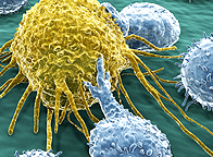
2023 Research: Exploratory Use of Extracellular Vesicles for CHRONIC Disorders
4/12/2023 - Dr. Paul Dreschnack, NY-based surgeon and biochemical researcher announced in a medical partnership for a new set of Extracellular Vesicles (EVs) research protocols. His research proposals are aimed to addressing some of the most highly challenging chronic disorders including PTSD, PSORIATIC ARTHRITIS, DIABETES, ALZHEIMER'S DISEASE, and AMYHOTROPHIC LATERAL SCLEROSIS. Advancements in areas of cell regenerative solutions include concepts in Extracellular Vesicles therapy which are now being explored for its potential as a natural product of our stem cells. Various in vitro studies have exhibited these nanoparticles or Extracellular Vesicles to contain advanced numbers of proteins, mRNA and lipids - all working to safely and effectively address the progression of specific given diseases. Though (at the time) no FDA regulatory clearances have been granted to the wide clinical use of EVs, continued research and clinical trials are underway to collect more data on EV applications and show substantial promise as novel treatment strategies for diseases. Launch plans for mid-2023 include IRB proposals, a national fundraising campaign and the submission to partner with specific development labs to conduct multi-validation medical studies of EVs. His new research A-team includes leaderships from the medical community including senior diagnostic imaging expert, Dr. Robert Bard (NYC), epigenetics and genomics expert Dr. Roberta Kline (NM), clinical psychiatrist Dr. Janice Greene (NYC), researcher Dr. Noelle Cutter and functional medicine practitioner Dr. Leslie Valle-Montoya (CA). - see complete article


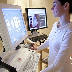
"SEEING IS BELIEVING": Advantages of Imaging in Research Studies

QUANTIFIABLE DETECTION OF THERAPY RESPONSE
MEDICAL IMAGING refers to the set of FDA approved modalities that are used to view the human body in order to diagnose and monitor medical conditions or the response of its treatments. The types of common modalities include X-rays, MRI (magnetic resonance imaging), Ultrasound, CT scan (computed tomography scan) and Nuclear medicine imaging including Positron Emission Tomography (PET) [1][2]
Ultrasound imaging (sonography) uses high-frequency sound waves to view inside the body. Because ultrasound images are captured in real-time, they can also show movement of the body's internal organs as well as blood flowing through the blood vessels. Unlike X-ray imaging, Ultrasound uses NO ionizing radiation exposure whatsoever.[3] This modality (sometimes referred to as "the orthopedic stethoscope"), offers easy access, portability, affordability and real-time monitoring and is safe for all patients, including those with cardiac pacemakers and metal implants, without any contraindications.

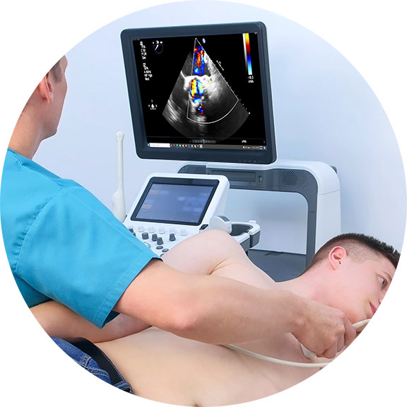
TRACKING THERAPEUTIC EFFECTS THROUGH BLOOD FLOW REACTIONS
To date, the ultrasound's ability to evaluate abnormalities within the soft tissue such as cysts, tumors and inflammation is used to help identify an expanded set of pathologies in the body.  Since the early 1970's, Dr. Robert L. Bard (NYC Cancer Radiologist) has used diagnostic imaging for pre and post procedural guidance, and diagnostic care (screening and monitoring) of his patients.  Dr. Bard is also recognized for his use of ultrasound in pharmaceutical research and clinical trials, where his leadership in analytical interpretation is sought after worldwide for identifying markers and therapeutic efficacy. 

Throughout his career, Dr. Bard has employed this imaging strategy to detect, track and confirm the body's reaction to a variety of therapeutic interventions. He has conducted medical center based double-blinded, corporate sponsored and private studies reviewing the effects of injectable therapies (PRP, Stem Cell therapies, etc) as well as non-invasive therapeutic interventions in studies of neuro-stimulation, electrostimulation and electromagnetic field treatments.  His approach involves the comparative study of measurable scanning data or quantitative ultrasound (QUS) which aims at recording interactions between the behavior and activity of biological tissue microstructure and ultrasound waves [5][6]. From a time-based comparative study of the treated area (before and after studies), Dr. Bard applies the use of blood flow detection technology or hemodynamic data gathering protocols, document specific objective and quantifiable biological responses to therapeutic treatments.

Ref:
1) Medical Imaging/FDA: https://www.fda.gov/radiation-emitting-products/radiation-emitting-products-and-procedures/medical-imaging#:~:text=Medical%20imaging%20refers%20to%20several,monitor%2C%20or%20treat%20medical%20conditions.
2) https://sonosimaging.com/press-corner/understanding-the-different-types-of-imaging/
3) https://www.fda.gov/radiation-emitting-products/medical-imaging/ultrasound-imaging
4) Diagnostic imaging to detect and evaluate response to therapy in bone metastases from prostate cancer: current modalities and new horizons https://pubmed.ncbi.nlm.nih.gov/26956538/


"Before & After" Studies

The most sensible and logical way to identify the results of any treatment is by tracking the body's response to it. Controlled testing must show the patient's condition PRE and POST effects, where true data-finding is collecting the necessary EVIDENCE of its claims. The investigator can pull a significant amount of data from this form of oberservational testing and recording: including stage-by-stage bodily response to future projections of possible side effects. Recording of any and all psysiological response means the researchers are counting on the patient's body to tell us what it is undergoing during the testing phase. To prevent mis-reading and erroneous reports, trials tend to work with a large number of test patients (commonly 50-100) and may also employ redundancies like undergoing multiple testing protocols for a second or even third opinion. To capture the benefits of a BEFORE AND AFTER review, Imaging is often used as a standard screening solution for the response of most of the major organs.


COMBINING MULTIPLE INNOVATIONS FOR IMPROVED WELLNESS
Today's clinical leaders are in constant search for new solutions to old problems. Historically, the medical community has uncovered many remarkable new solutions to countless health issues and symptoms by combining the use of compatible innovations. The AngioFoundation supports the test-driving & evaluation of current therapeutic solutions and exploring the concept of joint application of multiple technologies to advance the prospect of improved therapeutic performance. By joining specific technologies that support common or complementary targeting, we may actually discover a new treatment paradigm or add significant response (including the reduction in recovery time) in the restorative process.

INDEPENDENT CASE STUDIES: EFFICACY & PROGRESS TRACKING
Use of Advanced Digital 3D Doppler Sonographic Imaging technology to MONITOR, TRACK, SCREEN and IDENTIFY all treatment responses, successes & efficacy. The AngioFoundation team is experienced with designing a comprehensive Clinical Trial format for all medical protocol developers. The Doppler system is the safest repeat scanning solution for performance monitoring and staging use (no radiation) allowing the tester to accurately achieve and generate a comparative view of Before-After studies. Working with all test patients within this proven and stable paradigm can be incorporated with other current imaging technologies such as MRI, CT etc. Acquire useful data from concurrent patient testing programs such as: duration of desired response, resistance analysis, dosage-effect ratios, statistics assessment, secondary physiological responses and more.
   
REPORTING FOR INSTITUTIONAL STANDARDS & COMPLIANCE
Dr. Bard has supported countless projects, many of them pertaining to the achievement of regulatory standards. From medical device companies, clinical labs and pharma products, our resarch programs have helped to achieve proper certifications and compliance. By this, we perform a complete and highly detailed objective analysis of the product in question- reviewing all available benefits, a comprehensive assessment of their intended usage and short and long term effects- detailed within your compliance applications, supportive documentation and ansilary reporting.
   
CUSTOMIZED DESIGNS OF RESEARCH STUDIES
As a researcher, Dr. Bard welcomes collaborating with all developers under varying types of research paradigms. To determine a conclusive study within sensible variable scenarios or to help identify and confirm the developer's intended results of their product design, we can offer a wide array of clincal trial design angles including: CASE-CONTROL STUDIES, CASE REPORTING, META ANALYSES, COST-BENEFIT ANALYSIS, DOUBLE BLIND STUDIES, EMPIRICAL RESEARCH just to name a few.


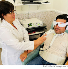
DEDICATED MONITORING OF PRE AND POST-TREATMENT PROGRESS
Within a closed testing program of 45-50 patients, a comparative review of pre and post treatment allows the developer a clear view of its efficacy and performance. Through the use of various imaging technologies and the experienced assessment strategies of our diagnosticians, we are able to provide the desired data which leads the devleoper to identify quality standards as they apply to the inevitable end user en masse.


Educational PROGRAMS
The AngioFoundation has been recognized worldwide by official medical organizations and peer reviews for its work in FIVE MAIN focal topics and class divisions. We continually maintain and update research works in these categories to support the advancement of these disciplines as part of The AngioFoundation's commitment to the scientific community.

Advanced use of Doppler Sonographic Imaging technology to identify malignant cancers and monitor their behavior through blood flow parameters- (Breast, Lung, Bladder, Prostate, Melanoma, etc) which correlates with comparative studies with other current technologies such as MRI, CT etc. 
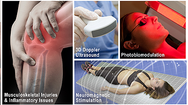
Research / assessment of musculoskeletal disorders (arthritis, inflammation, trauma) and dermatological issues through the advanced use of 3D/4D ULTRASOUND innovations. TECH REVIEWER: Beta-testing, industry-wide comparative feature review/evaluation program. Drafting of FDA application / compliance documentation of digital imaging technologies (models, brands and generations) including sub-dermal and musculoskeletal treatment devices Internal study of all cancer issues and health disorders of victims associated with 9/11 and other disaster-related environmental toxic exposures. Collaboration with geological labs & environmental statistics. (See First Responders Cancer Resource) 
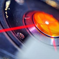
Function review / performance evaluation program of all laser-based medical equipment including devices specializing in sub-dermal musculatory treatment of chronic disorders.


Research results in APPLIED SCIENCE

IMAGING ASSISTS SURGICAL PLANNING OF INDICATED BIOPSIES

In my extensive career as the medical director of an advanced imaging diagnostics practice, I have provided great assistance to many surgeons with my work using advanced Doppler Scanning of Tumors and Cosmetic Disorders. I have uncovered countless dermal and subcutaneous issues that would have otherwise gone undetected with less effective technologies, leading to potential complications in the surgical procedure and patient recovery. The advancement in this innovation empowers upcoming surgical procedures with remarkable confidence of a safer end result. Where biopsies are becoming a thing of the past, our non-invasive 4D Digital imaging replaces weeks of lab work and radiologic tests and often provides more useful information.

DIGITAL BIOPSY CASES: WHAT ARE YOU ABOUT TO BIOPSY? WHAT HAPPENS AFTER THE NEEDLE INSERTS? Here we have 2 subdermal masses which are non tender and firm with no history of trauma. Case A: The oval mass (dark echoes=suspicious) with irregular vessels (red) was referred as a probable cyst or lipoma. The tumor is highly vascular and connected from the aorta by way of the subclavian feeding artery. Liposuction could result in massive hemorrhage and spread of tumor cells into the circulation.


Case B: The ovoid white region (bright echoes=benign) is ossified as confirmed by the CT scan of the coccyx. The sonogram allows you to reassure the patient it is NOT CANCER. It prompts one to avoid a standard needle that could bend, crack or dislodge into the soft tissues requiring further exploration to locate/retrieve the broken metal fragment.