 |
||||
| The Angio Institute | Research & Education | Brain Health Program | AngioNews/Media | Joining Hands |
|
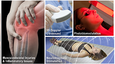
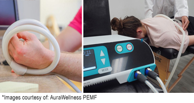
WAGING "WAR AGAINST INFLAMMATION" Refs: • Demo Day 1 (ASPEN LASER) | • Science behind THERALIGHT? | • PEMF 101 |
||||||||||||
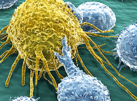
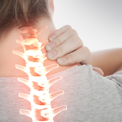
INFLAMMATION 101 The five cardinal signs are heat, pain, redness, swelling, and loss of function. Inflammation is a generic response, and therefore it is considered as a mechanism of innate immunity. Too little inflammation could lead to progressive tissue destruction by the harmful stimulus (e.g. bacteria) and compromise the survival of the organism. In contrast, too much inflammation, in the form of chronic inflammation, is associated with various diseases, such as hay fever, periodontal disease, atherosclerosis, and osteoarthritis. ACUTE inflammation is the initial response of the body to harmful stimuli from the blood into the injured tissues. A series of biochemical events propagates and matures the inflammatory response within the injured tissue. Prolonged or CHRONIC inflammation, leads to a progressive shift in the type of cells in the inflamed area and is characterized by simultaneous destruction & healing of the tissue from the inflammatory process.
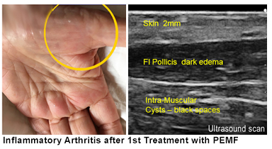
QUANTITATIVE IMAGING CONFIRMS THERAPEUTIC RESPONSE
Advancements in ENERGY MEDICINE - By: Dr. Roberta Kline
PHOTOBIOMODULATION
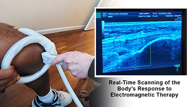
ELECTROMAGNETICS This educational literature is brought to you by the educational dept. of: BARD DIAGNOSTIC IMAGING |
|||||||||||||
 |
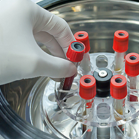
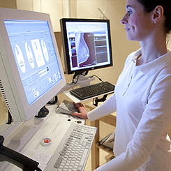
CLINICAL RESEARCH PROGRAM: DEVICE VALIDATION THROUGH IMAGING 11/25/2022- The concept behind ‘MEDICAL VALIDATION’ underscores the commitment of “ensuring that health or medical devices shall consistently provide the effects and benefits they are intended. Dr. Robert Bard (NYC) officially drafted an official update to his blueprint coordinating clinical monitoring and validation of non-invasive devices through diagnostic imaging protocols (under Institutional Review Board approved through HHS and FDA regulations). The primary monitoring and biometric reporting research protocol employs the use of medical grade 3D Doppler Ultrasound imaging ... (PDF:see details) |
||||||||||||
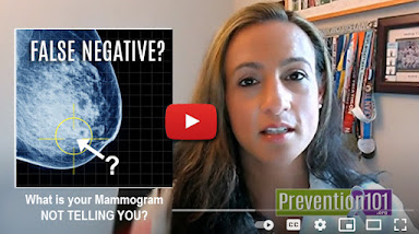 |
RESEARCH PROJECT: DENSE BREAST IN ATHLETIC COMMUNITY- 9/22/2021- For women with any level of breast density, one of the major concerns is the alarming rate of false positives that may align with cancers missed by a mammogram. The other concern is that women with dense breasts have a naturally higher risk of breast cancer than women with fatty breasts, and the risk increases with increasing breast density. (This increased risk is separate from the effect of dense breasts on the ability to read a mammogram.). The main focal points of this research project covers the diagnostic study of ATHLETIC WOMEN or those with LOW BODY MASS INDEX (bet 12-22% body fat). |
||||||||||||
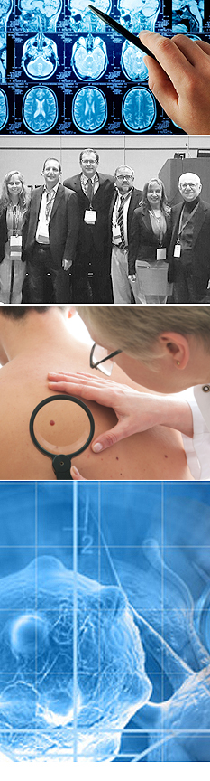
"SEEING IS BELIEVING": Advantages of Imaging in Research Studies
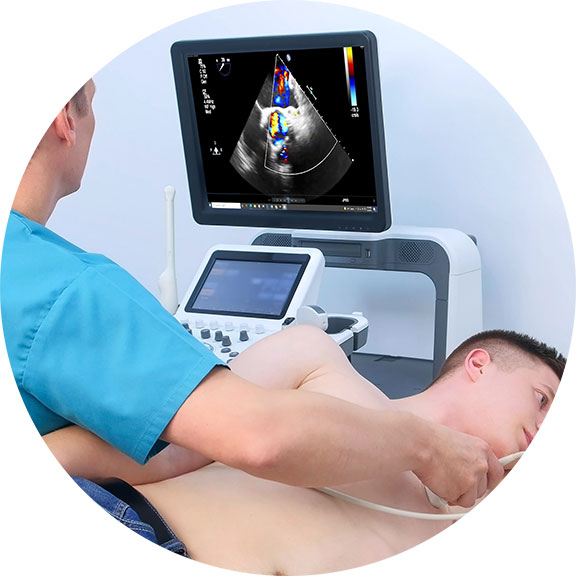
TRACKING THERAPEUTIC EFFECTS THROUGH BLOOD FLOW REACTIONS Throughout his career, Dr. Bard has employed this imaging strategy to detect, track and confirm the body's reaction to a variety of therapeutic interventions. He has conducted medical center based double-blinded, corporate sponsored and private studies reviewing the effects of injectable therapies (PRP, Stem Cell therapies, etc) as well as non-invasive therapeutic interventions in studies of neuro-stimulation, electrostimulation and electromagnetic field treatments. His approach involves the comparative study of measurable scanning data or quantitative ultrasound (QUS) which aims at recording interactions between the behavior and activity of biological tissue microstructure and ultrasound waves [5][6]. From a time-based comparative study of the treated area (before and after studies), Dr. Bard applies the use of blood flow detection technology or hemodynamic data gathering protocols, document specific objective and quantifiable biological responses to therapeutic treatments. Ref: |
|||||||||||||
"Before & After" Studies The most sensible and logical way to identify the results of any treatment is by tracking the body's response to it. Controlled testing must show the patient's condition PRE and POST effects, where true data-finding is collecting the necessary EVIDENCE of its claims. The investigator can pull a significant amount of data from this form of oberservational testing and recording: including stage-by-stage bodily response to future projections of possible side effects. Recording of any and all psysiological response means the researchers are counting on the patient's body to tell us what it is undergoing during the testing phase. To prevent mis-reading and erroneous reports, trials tend to work with a large number of test patients (commonly 50-100) and may also employ redundancies like undergoing multiple testing protocols for a second or even third opinion. To capture the benefits of a BEFORE AND AFTER review, Imaging is often used as a standard screening solution for the response of most of the major organs.
|
|||||||||||||
|
|||||||||||||
 QUANTIFIABLE DETECTION OF THERAPY RESPONSE
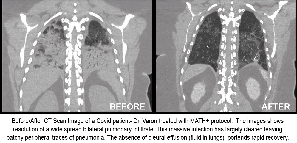


| DEDICATED MONITORING OF PRE AND POST-TREATMENT PROGRESS Within a closed testing program of 45-50 patients, a comparative review of pre and post treatment allows the developer a clear view of its efficacy and performance. Through the use of various imaging technologies and the experienced assessment strategies of our diagnosticians, we are able to provide the desired data which leads the devleoper to identify quality standards as they apply to the inevitable end user en masse. |
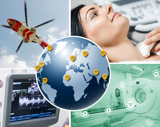
From Local to Global: The subdermal ultrasonic technology has become a powerful new standard in diagnosing and treating patients. The movement to "go digital' also brings significant advantages between clinicians in different locations by allowing them to share patient scans and test images in real time. This benefit is also greatly used in clinical trials from multiple locations. Working with a team is no longer limited to localized personnel; today's medical project allows complete working access to the most credentialed experts from any part of the globe. Dr. Bard has proven this work paradigm throughout his career -having partnered with countless clinical teams and research groups in European medical centers who sought his specific experience and diagnostic talents. Thanks to the use of the latest CLOUD-BASED imaging programs, trial projects can easily acquire, host, transfer and securely deliver any and all 3D/4D subdermal imaging of a patient (or test case) via REMOTE ACCESS. This innovation significantly adds a new level of efficiency and performance to any research project while greatly reducing trial costs and delays. |
|||
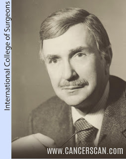
Contributing to the evolution of SCIENCE by: Dr. Robert L. Bard
I welcome all solutionists and innovators who support the healing arts community. I have devoted my specialized work to pursue the advancement of MEDICAL RESEARCH with NON-INVASIVE SURROGATE ENDPOINTS with the hopes of contributing my talents to the performance of your current and future projects. With over 45 years in the field of advanced diagnostic science, my life's work has been about the clinical examination and targeted analyses of all subdermal disorders using the latest quantifiable digital imaging innovations. I have established an entire foundation dedicated to Medical Research committed originally to the exploratory studies of all cancer treatment proctocols. Alongside this, I have also been most active in collaboration with some of the top treatment strategists, health centers, clinical labs, experimental / alternative treatment professionals and medical device manufacturers. I have earned a reputation for my investigative approach within various examination paradigms including progress monitoring and surveillance. As a CO-INVESTIGATOR, I have the greatest interest in supporting all entities committed to contributing technical innovations and new advancements in treatment solutions for our medical community. By this, I wish to build a partnership with establishing research teams to conduct clinical test projects where my talents to support visual reporting through imaging to be a priceless benefit to your overall objectives. Most research sponsors and medical developers' testing and tracking needs often fall into one of a number of common task categories- all within the objectives of public health, safety and healthcare support. I look forward to learning about your project with the hopes of supporting your needs and objectives. |
|||
Research & Educational PROGRAMS • Efficacy of Injected Hyaluronidase Filler Reduction Dept Dermatology Mt Sinai • Photoacoustic and Optical Imaging • Doppler Evaluation of Radiation Treatment Response • 3D Doppler Sonography of Malignant Melanoma • 3D Imaging of Psoriasis • Doppler Vessel Density in Melanoma • Prostatic Volume Reduction Implementing Radiotherapy Treatment Application • Ultrasonography of the Spine in Rheumatoid Arthritis • Ultrasound Evaluation of the Patient with Rheumatoid Arthritis Click HERE at pgs 19-21 for a complete list of Research Projects |
|||
| `
Scientific PUBLICATIONS (see Pg. 6-19 books published & international presentations) |
|||
Recent & Innovative PROJECTS Dr. Bard's 4D Doppler capabilities have become the preferred non-invasive solution with research programs to seek out treatment testing or application performance tracking for this exact type of exploratory testing. Dr. Bard has the capacity to identify and track prestroke vasospasms, any increase in intracranial pressure, brain aneurysms and many degenerative disorders. (See related article) |
|||
Early collaboration with technological FRONTIERSHIP
In 1976, during Dr. Bard's days as a young radiologist, he was approached by Dr. Henry Leis Jr., the pioneer doctor who wrote the very first text on breast cancer and developed mammography 18- a means of early diagnosis and instrumental in the use of many of the less invasive procedures used in the treatment of breast cancer today. He confessed with great concern that he had all these patients with lumpy or cystic breasts developing tumors that he could clearly feel but the mammogram kept missing it. Seeking Dr. Bard's help through sonogram technology, they worked on his patients together and the sonogram clearly identified and quickly diagnosed a mass as either a cancer or a benign cyst, in a dense, lumpy breast. Since then, the sonogram became incorporated in high-risk patients’ regimen every six months religiously because it finds tumors while they're small and “lumpectomy” surgery is curative if the mass is less than 1 cm. This is alongside doing mammograms once a year in women over 50 or unless they have a history of cancer- at which case, we do it starting at age 45. |
|||
Research Support and Fundraising EVENTS
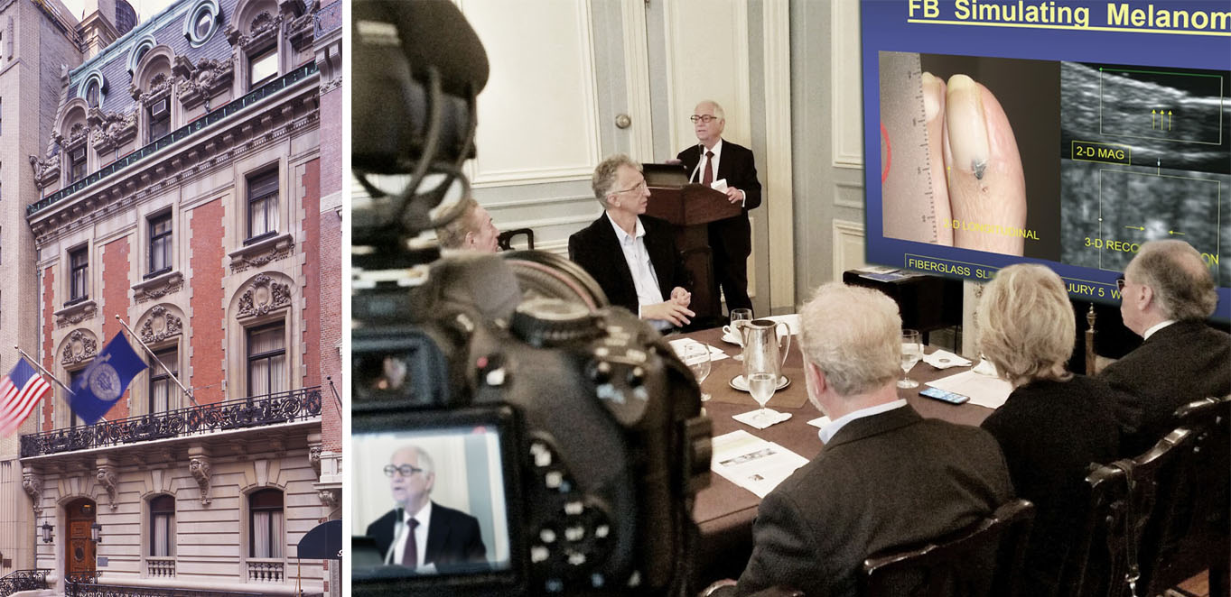
May 10, 2018- NYC; The LOTOS CLUB hosted the 2018 speaker series with Dr. Robert Bard's opening presentation on Early Detection & Prevention by NYCRA (New York Cancer Resource Alliance) and endorsed Dr. Bard to launch this theme because of his expanded knowledge in today's modern diagnostic technologies and non-invasive applications including the 4D Doppler ultrasound technology - much of which has been driven by European medical influence for the cancer treatment community. Dr. Bard is an internationally recognized leading expert in the use of this process for the screening, scanning and pre/post procedural monitoring of Breast, Prostate & Skin Cancers. He is also known for his public mission to replace surgical analysis of tumors with his coined "digital biopsies". Dr. Bard gave the standing-room only audience at the LOTOS CLUB an astounding look at the effectiveness of his imaging process to the surgical chain. He shared images of actual patient cases including the study of blood flow in tumors, behaviors of cysts, foreign bodies in trauma cases, fracture analysis and proven ways that cancers can be safely detected, studied and even treated with the use of non-surgical means. The high accuracy of 3D-4D Histogram analysis allows cancer treatment to be implemented without invasive biopsies that may spread tumor cells. (full article) |
|||
Educational PROGRAMS
The AngioFoundation has been recognized worldwide by official medical organizations and peer reviews for its work in FIVE MAIN focal topics and class divisions. We continually maintain and update research works in these categories to support the advancement of these disciplines as part of The AngioFoundation's commitment to the scientific community.
 Advanced use of Doppler Sonographic Imaging technology to identify malignant cancers and monitor their behavior through blood flow parameters- (Breast, Lung, Bladder, Prostate, Melanoma, etc) which correlates with comparative studies with other current technologies such as MRI, CT etc. Advanced use of Doppler Sonographic Imaging technology to identify malignant cancers and monitor their behavior through blood flow parameters- (Breast, Lung, Bladder, Prostate, Melanoma, etc) which correlates with comparative studies with other current technologies such as MRI, CT etc. |
 Research / assessment of musculoskeletal disorders (arthritis, inflammation, trauma) and dermatological issues through the advanced use of 3D/4D ULTRASOUND innovations. Research / assessment of musculoskeletal disorders (arthritis, inflammation, trauma) and dermatological issues through the advanced use of 3D/4D ULTRASOUND innovations. |
 TECH REVIEWER: Beta-testing, industry-wide comparative feature review/evaluation program. Drafting of FDA application / compliance documentation of digital imaging technologies (models, brands and generations) including sub-dermal and musculoskeletal treatment devices TECH REVIEWER: Beta-testing, industry-wide comparative feature review/evaluation program. Drafting of FDA application / compliance documentation of digital imaging technologies (models, brands and generations) including sub-dermal and musculoskeletal treatment devices |
 Internal study of all cancer issues and health disorders of victims associated with 9/11 and other disaster-related environmental toxic exposures. Collaboration with geological labs & environmental statistics. (See First Responders Cancer Resource) Internal study of all cancer issues and health disorders of victims associated with 9/11 and other disaster-related environmental toxic exposures. Collaboration with geological labs & environmental statistics. (See First Responders Cancer Resource) |
 Function review / performance evaluation program of all laser-based medical equipment including devices specializing in sub-dermal musculatory treatment of chronic disorders. Function review / performance evaluation program of all laser-based medical equipment including devices specializing in sub-dermal musculatory treatment of chronic disorders. |